MCL LCL PCL
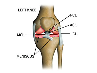
These are the 4 main ligaments or tough bands of tissue in the knee. ACL is anterior cruciate ligament which connects the thigh bone to the shin bone. MCL is another of the four ligaments, the medical collateral ligament.
Medial Collateral Ligament (MCL) Tear
What is a MCL Injury?
The medial collateral ligament (MCL) runs from the inside surface of the upper shin bone to the inner surface of the bottom thigh bone. This ligament keeps your shin bone (tibia) in place.
The MCL is usually injured by pressure or stress on the outside part of the knee. A block to the outside part of the knee during football is a common way for this ligament to be injured.
An MCL injury can be a stretch, partial tear, or complete tear of the ligament. MCL injuries also often occur at the same time as an anterior cruciate ligament (ACL) injury.
Common symptoms of an injury to the medial collateral ligament are:
- Knee swelling
- Locking or catching of the knee when you move it
- Pain and tenderness along the inside of the joint
- A knee that gives way or feels like it is going to give when it is active or stressed in a certain way
If you are being treated for an MCL injury or were recently, be sure to contact your health care provider if you notice:
- Increased instability in your knee
- Pain or swelling after they initially faded
- That your injury is not getting better with time
- You re-injure your knee
Lateral Collateral Ligament (LCL) Tear
What are LCL tears?
A lateral collateral ligament (LCL) tear is a knee injury that causes pain, swelling and bruising. Your LCL is a band of tissue located on the outside of your knee (the side that faces away from your body). This tissue connects your lower leg bones to your thigh bone. It stops your knee from bending outward abnormally.
Athletes in sports like football, soccer and skiing are at higher risk for LCL tears, which can prevent you from competing. However, with time, treatment and rehabilitation, you should be able to play some sports again.
How does the knee usually work?
Three bones make up your knee joint:
- Your thighbone (femur)
- Your kneecap (patella)
- Your shinbone (tibia)
Ligaments hold the bones together. There are two types:
- Collateral ligaments: These ligaments are on the sides of your knee. The medial (inside) ligament connects your femur and tibia. The lateral (outside) collateral ligament (your LCL) connects the femur and fibula. Thanks to these ligaments, you can move your knee sideways.
- Cruciate ligaments: Your cruciate ligaments are inside your knee joint. They cross each other (the anterior cruciate ligament is in the front and the posterior cruciate ligament is in the back) and they form an “X.” These ligaments control the way your knee moves back and forth.
What are the symptoms of LCL tears?
The symptoms of an LCL tear include:
- Pain
- Swelling
- Tenderness
- Bruising
- An unstable feeling. Your knee might feel like it’s about to give out or buckle or lock up.
You might find that the sensation of being unstable on your feet continues after you’re walking again. While not unusual, it’s a good idea to tell your healthcare provider about it. Such instability can feel a little scary since you might fear injuring yourself again.
How are LCL tears diagnosed?
When you see your healthcare provider (which you should do as soon can), they’ll ask you questions about your injury and look at your knee. They’ll check for the following:
- Tenderness
- Swelling
- How your knee moves
- How your leg moves
- Other injuries
Your healthcare provider might order tests, including:
- X-ray
- MRI
- Ultrasound
posterior cruciate ligament (PCL)
What is a PCL Injury?
The posterior cruciate ligament (PCL) is the strongest ligament in the knee. It extends from the top-rear surface of the tibia (bone between the knee and ankle) to the bottom-front surface of the femur (bone that extends from the pelvis to the knee). This ligament prevents the tibia from moving too much and going behind the femur.
A PCL injury (which may also be called hyperextended knee) is a partial or complete tearing or stretching of any part of the posterior cruciate ligament. The PCL is usually injured by overextending the knee (hyperextension). This can happen if you land awkwardly after jumping.
The PCL can also become injured from a direct blow to the flexed knee, such as falling hard on a bent knee.
Most PCL injuries occur with other ligament injuries and severe knee trauma. Often, the knee is dislocated and the nerves and blood vessels are injured.
If you suspect PCL injury, it is important to be seen by a doctor right away. PCL injuries often occur with other ligament injuries or severe knee trauma, so you should be checked early for these other conditions.
- The symptoms of a PCL injury include:
- Knee swelling and tenderness in the space behind the knee (popliteal fossa)
- Knee joint instability
- Knee joint pain
Diagnosis of PCL Injury
If you or your doctor suspect a PCL injury, your health care provider will perform a physical examination to check for signs of PCL injury. Your doctor will begin by moving the knee joint in various directions.
Your doctor may also check if there is fluid in your knee joint. This test may show bleeding into the joint.
Other tests that may be ordered include:
- Knee MRI
- Knee joint x-ray